Gastroscopy & Upper Endoscopy
When you have unexplained stomach issues, you may need an upper endoscopy. Call today to schedule an appointment with Dr. Yanez Peerbaccus or go online to book your initial consultation.
What is a gastroscopy/upper endoscopy?
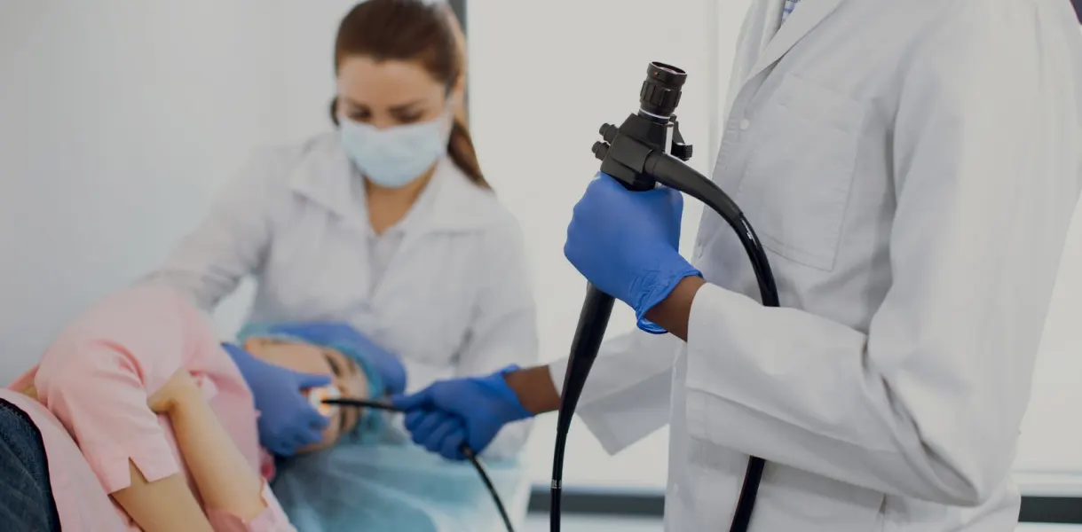
A gastroscopy/upper endoscopy, sometimes called OGD, is a medical procedure that utilises an advanced medical instrument known as an endoscope. This device is characteristically long and flexible, featuring a cutting-edge video camera affixed to its tip, which provides real-time, high-definition images of the internal structures of the gastrointestinal (GI) tract. The endoscope is gently inserted through the patient’s mouth and meticulously navigated through the esophagus into the stomach and duodenum, the initial portion of the small intestine. The primary objective of this procedure is to afford the physician an up-close view of these areas, facilitating a thorough examination. Furthermore, gastroscopy is versatile, enabling the doctor to conduct specialised interventions, such as the extraction of tissue samples for biopsy, directly through the endoscope. Typically, gastroscopy is performed on an outpatient basis within a hospital setting.
Who Needs an Upper Endoscopy?
Your gastroenterologist may recommend an upper endoscopy if you have unexplained gastrointestinal issues like:
Chronic or acute abdominal pain, persistent indigestion, unexplained nausea, frequent episodes of vomiting, and difficulties in swallowing. It is notably the most reliable technique for identifying the origins of bleeding within the upper GI tract, significantly outperforming conventional imaging methods like x-rays in detecting conditions such as inflammation, ulcers, or tumours.
A gastroscopy is invaluable for tissue sampling, which is essential for the diagnosis of a range of conditions. Through biopsies obtained during the procedure, it is possible to detect sites of infection, evaluate the functional state of the small intestine, and diagnose abnormal tissue growths, including malignant lesions and conditions like coeliac disease.
The procedure allows for the introduction of miniature surgical tools through the endoscope, enabling physicians to treat detected abnormalities with minimal discomfort to the patient. These therapeutic interventions can vary widely, from dilating constricted areas within the GI tract, removing polyps, which are generally benign growths, to employing techniques to stop gastrointestinal bleeding.


What is the preparation for an upper endoscopy/ gastroscopy?
Preparatory measures for gastroscopy are critical to the procedure’s success and the patient’s safety. To ensure a clear view of the GI tract, it is imperative that the stomach be completely empty. Patients are thus advised to abstain from eating or drinking, including water, for approximately six hours before the procedure. Specific instructions regarding preparation will
be tailored to the individual patient, depending on the scheduled time for the gastroscopy, and will be provided by your doctor.
Patients scheduled for gastroscopy should engage in detailed discussions with their physician about their current medications. Adjustments may be necessary, especially for those who take diabetic medications and blood thinners. Additionally, it is crucial to disclose any allergies to medications and inform the physician about existing medical conditions, particularly those affecting the heart or lungs, to ensure a personalised and secure approach to conducting the gastroscopy.
How is an upper endoscopy/gastroscopy performed?
The process of undergoing a gastroscopy begins with the administration of deep sedation, designed to induce a state of relaxation without the complete unconsciousness associated with a general anesthetic. This means that while you might have a very faint awareness of the surrounding activities, it’s unlikely that you’ll retain any memory of the procedure. To minimise discomfort and suppress the gag reflex, the back of your throat may be sprayed sometimes with a local anesthetic, rendering it numb. Additionally, to protect your teeth as well as the endoscope, a small, protective mouthguard is placed between your teeth. For those who wear dentures, these will need to be removed prior to the procedure.
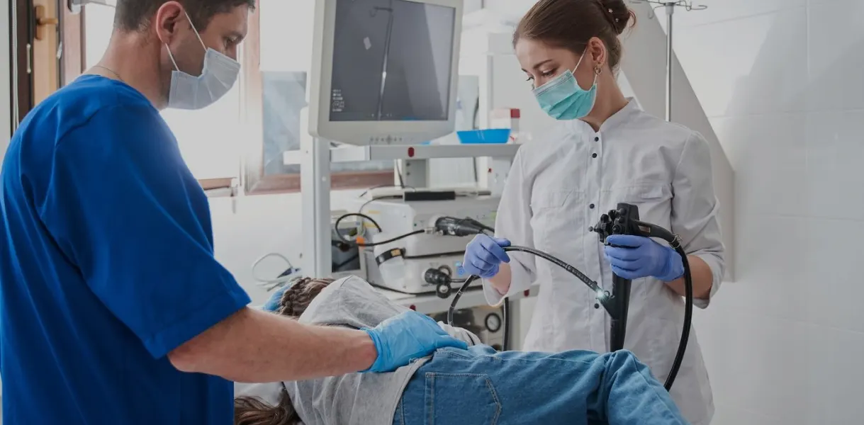
The gastroscopy itself is relatively brief, typically lasting between 10 to 20 minutes. During this time, you will be positioned comfortably on your left side. The endoscope, a slender tube about less than one centimeter in diameter, is gently introduced into the mouth and carefully advanced through the esophagus into the stomach and duodenum, the initial segment of the small intestine. It’s important to note that the endoscope does not obstruct the windpipe, so your ability to breathe remains unaffected.
A sophisticated miniature camera situated at the tip of the endoscope sends real-time video to a monitor, enabling the physician to conduct a detailed inspection of the lining of your upper gastrointestinal tract. This thorough examination allows for the identification of any abnormalities or signs of conditions that could be causing symptoms or require further intervention.
What happens after the procedure is completed?
After your gastroscopy, you’ll be taken to a designated recovery zone where healthcare professionals will monitor you until the sedatives’ effects significantly decrease. During this period, you may experience some soreness in the throat, this discomfort results from the endoscope’s passage. Additionally, you might feel unusually bloated; this sensation is due to the air that was introduced into your stomach during the procedure to enhance visibility and facilitate a thorough examination.
Typically, you are permitted to resume eating after you depart from the medical facility, unless your doctor provides specific dietary restrictions or recommendations based on the findings of your gastroscopy.
Shortly after the procedure, your doctor will likely offer a preliminary overview of the findings. This initial assessment is valuable, but it’s important to remember that a more
comprehensive discussion is usually reserved for a subsequent follow-up appointment. This is especially true if biopsies were taken or samples collected for laboratory analysis, as these processes require several days to complete.
Due to the sedatives administered for your comfort during the gastroscopy, it’s imperative to avoid certain activities for a period of 24 hours. These activities include driving vehicles, using public transportation without accompaniment, operating any machinery, signing legally binding documents, or consuming alcoholic beverages. It’s highly advisable to have a friend or relative escort you home and remain with you for some time as a precautionary measure to ensure your safety until the sedative’s effects have completely worn off. Full recovery from the procedure is typically expected by the following day after about 24 hours. Paying close attention to and adhering to any discharge instructions provided is crucial for a smooth recovery.
What are the possible complications of the procedure?
Potential complications, though rare, include a mildly sore throat post-procedure, a slight risk of chest infection, bloating due to air trapped in the stomach, or minor bleeding if a biopsy was performed or specific treatments were administered during the procedure. An extremely rare yet serious complication is the perforation or tearing of the stomach lining, necessitating immediate hospital admission and surgical repair. Additionally, adverse reactions to the sedative used, though uncommon, are possible.
If the gastroscopy does not conclusively address the reason for the procedure or cannot be completed as planned, a repeat procedure might be necessary. Should you experience any of the following symptoms in the hours or days following your gastroscopy such as fever, difficulty swallowing, increasing pain in the throat, chest, or abdomen, or any other symptoms that raise concern, it’s critical to immediately reach out to your specialist or the hospital for guidance and further evaluation.
What is an oesophageal dilatation?
The esophagus, commonly known as the food pipe, is the muscular tube responsible for transporting food from the mouth to the stomach. It accomplishes this by contracting its muscles in a coordinated manner, propelling food downward. However, there are circumstances where the esophagus can become constricted or narrowed, complicating the process of swallowing and making the intake of food challenging.
Such narrowing, medically referred to as stricture, can arise due to several reasons. A common cause is the formation of scar tissue resulting from surgical procedures performed on the esophagus, from reflux oesophagitis and eosinophilic oesophagitis. Another potential cause is injury from the ingestion of caustic substances, leading to burns inside the esophagus.
To diagnose this narrowing, healthcare providers often resort to a diagnostic test known as a barium swallow study. This procedure requires the patient to drink a barium-containing liquid, which is designed to coat the lining of the esophagus and stomach, making them visible on X-ray images. As the barium moves down the esophagus to the stomach, X-rays are taken at various intervals to capture the passage of the liquid and highlight any areas of
narrowing or obstruction. Alternatively, a stricture can be diagnosed while performing an upper endoscopy.
If a stricture is identified, one therapeutic option available is esophageal dilatation. This procedure is aimed at widening the constricted segment of the esophagus to alleviate symptoms and improve the patient’s ability to swallow. In preparation for this procedure, it’s common for doctors to request a barium swallow study to precisely locate the narrowed area and to gather detailed information about its extent and severity. This preparatory step is crucial for planning the dilatation procedure effectively and ensuring its success.
How is oesophageal dilatation performed?
Once the narrowed portion is identified, the doctor selects an appropriate dilating device based on the size and location of the narrowing. There are several types of dilating devices used, including balloons and bougies (tapered dilators).
In balloon dilation, a deflated balloon attached to the end of the endoscope is positioned at the site of the narrowing. The balloon is then inflated to stretch the narrowed area. The pressure and duration of inflation are carefully controlled to avoid over-dilation and minimise the risk of complications.
Bougie dilation involves using a series of progressively larger bougies (tapered dilators) to gradually stretch the narrowed area. The bougie is passed through the narrowed portion of the esophagus, incrementally increasing in size until the desired diameter is achieved.
Throughout the dilation procedure, the doctor monitors the patient’s vital signs and watches for any signs of complications such as bleeding or perforation of the esophagus.
After the dilation is completed, the endoscope is removed, and the patient is monitored for a period to ensure there are no immediate complications. They may experience some discomfort or soreness in the throat after the procedure. Depending on the underlying condition being treated, the patient may be advised to follow specific dietary or lifestyle recommendations to promote healing and prevent recurrence of the narrowing.
What are the possible complications of dilatation?
Overall, esophageal dilation is a relatively safe and effective procedure for treating narrowed segments of the esophagus, but it carries some risks, including bleeding, perforation, and recurrence of the narrowing. Patients should discuss the potential risks and benefits of the procedure with their healthcare provider before undergoing esophageal dilation.
